Wednesday, August 15, 2012
anterior cases
I made this veneer on #9 with emax LT block and E4D, stained and glazed it.
I normally send out all anterior cases, this one I gave it a try because #8 was a full porcelain crown made by previous dentist, and it seems pretty easy to match.
This second one is a replacement of PFM crown.
The last photo was taken of a try-in with glycerin, lab fabricated this empress crown. gingiva was still red due to lack of good oral hygiene.
somehow on this day my flash on left did not work, sorry about the shadow on the left.
The next one is 2 veneers and 2 crowns
Onlays as alternative to crown
I believe that we, dentist, cut too much sound tooth structure away on daily basis. "Large filling, no problem, we will put a crown on it." "Filling keeps coming off or breaking? let's put a crown on it."
"Broken tooth or filling? let's crown it"
Teeth are not like hair or nails, it does not grow back after cut.
I made this onlay (lava ultimate) with E4D, cemented in the same appointment.
Tooth #30 had a deep O composite, fracture lines on D and mid-lingual.
Occlusal composite removed, crack line at the base of DL cusp.
Caries detector used, and the finished restoration only covering DL cusp, and D marginal ridge.
Buccal cusps and ML cusp are all kept as untouched. Did not extend margins beyond old composite.
"Broken tooth or filling? let's crown it"
Teeth are not like hair or nails, it does not grow back after cut.
I made this onlay (lava ultimate) with E4D, cemented in the same appointment.
Tooth #30 had a deep O composite, fracture lines on D and mid-lingual.
Caries detector used, and the finished restoration only covering DL cusp, and D marginal ridge.
Buccal cusps and ML cusp are all kept as untouched. Did not extend margins beyond old composite.
Sunday, April 1, 2012
newly erupted molars
Now I used a cotton pellet ( as big as needed) as gingival retraction, to push away the gingiva and to control fluid. Laser gingivectomy can also be done, but most of time, I find the cotton pellet is sufficient.
I use it from the beginning, if burs are needed to remove caries, it will keep it away from getting touched by the bur. Sometimes the sandblasting is enough to remove decalcified enamel.
 |
| pre-op view with decay on occlusal, DB cusp covered |
 | ||
| cotton pellet on distal, caries removal started. |
 |
| composite placed and cured |
Friday, March 23, 2012
distal decay on 2nd molars.
I followed Pascal Magne's advice on this one, and it turned out really nice and easy. Check out the sequence:
 |
| pre-op taken 11/2011 |
 |
| decay exposed after #32 extracted |
 |
| matrix band placed, caries detector used |
 |
 |
| pulp capping and primer |
 |
| immediately after band removal, prior to finishing or polishing |
 |
| pot-op x-ray, no finishing or polishing on distal |
Cut a second piece and slide into the first band to even go deeper and to protect gingiva during caries removal. Discard it prior to placing composite. Voila!
If an indirect restoration (onlay) is needed on 2nd molar with distal decay, this technique can be used for margin elevation. check out the next case:
 |
| #18 MO composite DO amalgam with open margin on distal |
 |
| amalgam removed, found base |
 |
| base removed, found decay |
 |
| different angle to show the DL wall |
 |
| MO composite removed |
 |
| still removing more decay |
 |
| I was really dreading seeing pulp, did not want to end up in RCT |
 | ||
| fortuanately, no pulpal exposure! I placed a composite to elevate the margin, IDS done |
 |
| CAD/CAM onlay cemented with composite as cement |
 |
| immediately after cementation |
Next case the decay was smaller, 3rd molar was extracted, crown lengthening done, and I used distal access instead of occlusal access. it was deeper than it looked.
 |
| pre-op bite-wing |
 |
| after extraction of #32 |
 |
| clinical view after crown lengthening, I thought it will be small... |
 | ||
| Wrong, it was almost to pulp | 1 | 1 |
Friday, January 27, 2012
IDS ( immediate dentin sealing) protocol
-prep tooth according to biomimetic principles
-etch exposed dentin for no longer than 15 seconds.
-gently dry leaving moist dentin
-apply primer (optibond FL) with microbrush for 15 seconds, evaporate solvent. if needed, apply second coat.
-apply bonding agent (optibond FL) to exposed dentin. DO NOT BLOW THIN, I was doing so as I usually do with composite restorations, and when I use sandblaster during cementation, some of the bonding agent was removed and it caused post-op sensitivity
-light cure
-air block with glycerin
-light cure again.
--scrub sealed dentinal area with fine pumice on cotton pellet to remove remaining oxygen inhibiting layer. rinse off pumice
-remove bonding agent on enamel or margins by oscillating tips, or finishing burs. pack cords to protect gingiva prior to finishing if necessary. Pack wax to block out undercut where the impression material can get locked and tear upon removal, it usually happens at gingival embrasures with margins away from gingiva
-impression.
it may look complicated, but it does not take much longer. the bonding strength is greatly increased in comparison to delayed bonding at cementation, there are actually a lot of benefits. The main one for me is: no post-op sensitivity!
Thursday, January 26, 2012
today I am attending a seminar in IDEA facility, with Pascal Magne and Michael Magne. Love it! Eye opener. If you ever have a chance, take it!
http://www.ideausa.net/
What is it all about? if you have not heard about Pascal Magne, it is all about Biomimetic principles, copying the nature, saving every bit of tissue as possible. Some of concepts are contradicting to what I learned in school ( not that I am that old), and to what majority of dentist believe in.
Just to illustrate:
-immediate dentin sealing after prep, prior to impression taking.
- margin elevation technique. if there is a subgingival margin, elevate it with composite and then place margin of indirect restoration on the equi or supra-gingival area.
-preservation of pulp (vital, normal response)at all cost. Do not kill the pulp, it starts what he calls the "circle of death" for tooth: RCT tooth, crown, crown with post, fracture or failed tooth, extraction. Even with deep caries removal, create a absolute healthy tooth structure periphery, and at the pink deepest part, leave it undisturbed.
-bilaminar approach to treat teeth when there is need for facial and lingual restoration, e.g. erosion and wear cases: ultraconservative veneer on facial, composite on palatal to restore wear facet. Do not make a full crown prep.
-never cut for full crown, even with a pre-existing crown replacement, sometimes we can get away from full crown. Onlays, inlays, veneers, whatever it takes to not remove more than needed. we don't need full crowns for tooth after RCT, we don't need full crowns after a big amalgam to "wrap and protect" the tooth.
-CAD/CAM composite onlays: very conservative, it can be 1mm thin on occlusal. use it to restore posterior may be better than emax or zirconia. harder is not always better. how much do we have to cut for porcelain, emax or zirconia? much deeper than 1mm, they are not kind to opposing dentition, bonding is harder ( talk about bonding to zirconia!).
-there is a on going research for CAD/CAM composite abutment for implants, and results are very promising. bonds well, can be prepped at chairside ( veneer prep), it absorbs and flex like dentin.
I wish I can spread, intrigue and convert as many dentist so we can all think being minimally invasive... to conserve and appreciate what nature gave us, and not to be " serial tooth killer".
Wednesday, January 25, 2012
occlusal caries
I have heard unexpected responses from patients when I inform them of caries:
" but I don't feel anything..."
" really? where? how come I can't see or feel it?"
These pictures can really illustrate how a caries can fool all of us, even dentists.
" but I don't feel anything..."
" really? where? how come I can't see or feel it?"
These pictures can really illustrate how a caries can fool all of us, even dentists.
deceiving caries
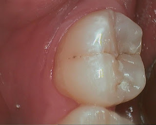
This tooth was diagnosed for occlusal composite over a year ago. Pt did not set appointment when I reminded him again that there is a large decay. He used demand force review system to question if the diagnosis was correct, because he had never had any symptoms, nor had been advised of so many filling before I took over previous owner's practice. BTW, demand force review system is a great way to get feedback from patients, there are a lot of information that they may feel uncomfortable sharing with you face to face. There is clearly a large radiolucency under the thick layer of occlusal enamel. The pre-op intra-oral photo does not show large decay, only deep grooves with a shadow coming through enamel.
I replied to him and promised that I can show him photos of the tooth while we are working on it, step-by-step. He came in for filling on #18. Here is a sequence of photos taken with caries detector:
I am pretty sure that it will very soon end up in a RCT if we have waited a little longer. it was huge as I kept removing decay.
I often hear people complaning how dentist drilled too much of their good tooth away, that a tiny hole can become a huge one. I think I/O pictures are the best tool to help them understand why we do what we do. It is also great documentation in case RCT was needed. Fortunately I did not have any pulpal exposure, but it was darn close! I was very glad that he came in, because there was no other way to prove to him how big the decay was, he took a leap of faith for allowing me to drill the tooth.
There are also interproximal caries on other teeth shown on bite-wing, I hope he will trust me after this one.
I replied to him and promised that I can show him photos of the tooth while we are working on it, step-by-step. He came in for filling on #18. Here is a sequence of photos taken with caries detector:
I often hear people complaning how dentist drilled too much of their good tooth away, that a tiny hole can become a huge one. I think I/O pictures are the best tool to help them understand why we do what we do. It is also great documentation in case RCT was needed. Fortunately I did not have any pulpal exposure, but it was darn close! I was very glad that he came in, because there was no other way to prove to him how big the decay was, he took a leap of faith for allowing me to drill the tooth.
There are also interproximal caries on other teeth shown on bite-wing, I hope he will trust me after this one.
Subscribe to:
Comments (Atom)


