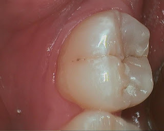
This tooth was diagnosed for occlusal composite over a year ago. Pt did not set appointment when I reminded him again that there is a large decay. He used demand force review system to question if the diagnosis was correct, because he had never had any symptoms, nor had been advised of so many filling before I took over previous owner's practice. BTW, demand force review system is a great way to get feedback from patients, there are a lot of information that they may feel uncomfortable sharing with you face to face. There is clearly a large radiolucency under the thick layer of occlusal enamel. The pre-op intra-oral photo does not show large decay, only deep grooves with a shadow coming through enamel.
I replied to him and promised that I can show him photos of the tooth while we are working on it, step-by-step. He came in for filling on #18. Here is a sequence of photos taken with caries detector:
I am pretty sure that it will very soon end up in a RCT if we have waited a little longer. it was huge as I kept removing decay.
I often hear people complaning how dentist drilled too much of their good tooth away, that a tiny hole can become a huge one. I think I/O pictures are the best tool to help them understand why we do what we do. It is also great documentation in case RCT was needed. Fortunately I did not have any pulpal exposure, but it was darn close! I was very glad that he came in, because there was no other way to prove to him how big the decay was, he took a leap of faith for allowing me to drill the tooth.
There are also interproximal caries on other teeth shown on bite-wing, I hope he will trust me after this one.


No comments:
Post a Comment